Cognitive Longevity
Brain Volume and Longevity:
What MRI Studies Actually Show
Brain scans increasingly get summarized as a single, striking number: "brain age." A person's actual age might be 55, but their scan might look like a 62-year-old's — or a 48-year-old's. This isn't just a novelty metric. A growing body of research uses this "brain age gap" to predict real outcomes, including all-cause mortality, with genuinely large effect sizes. This article covers what that metric actually measures, how strongly it predicts outcomes, and how hippocampal volume specifically functions as a diagnostic tool in the space between healthy aging and dementia.
Key numbers
| Finding | Detail |
|---|---|
| Mortality risk per 1-year increase in "brain age gap" | 12% higher all-cause mortality (HR 1.12) |
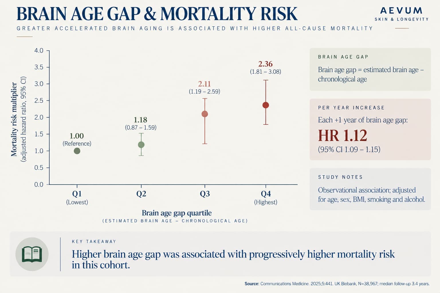
| Mortality risk, highest brain-age-gap quartile vs. lowest | 2.36x higher all-cause mortality |
| Alzheimer's disease risk per 1-year brain age gap increase | 16.5% higher |
| Study scope | UK Biobank (n=38,967), ADNI (n=1,402), PPMI (n=1,182) — multi-cohort validation |
| Hippocampal volumetry diagnostic accuracy, AD vs. healthy | 82% sensitivity / 87% specificity (33 studies, 5,157 patients) |
| Hippocampal volumetry diagnostic accuracy, MCI vs. healthy | 60% sensitivity / 75% specificity — notably weaker at the earlier stage |
How it works
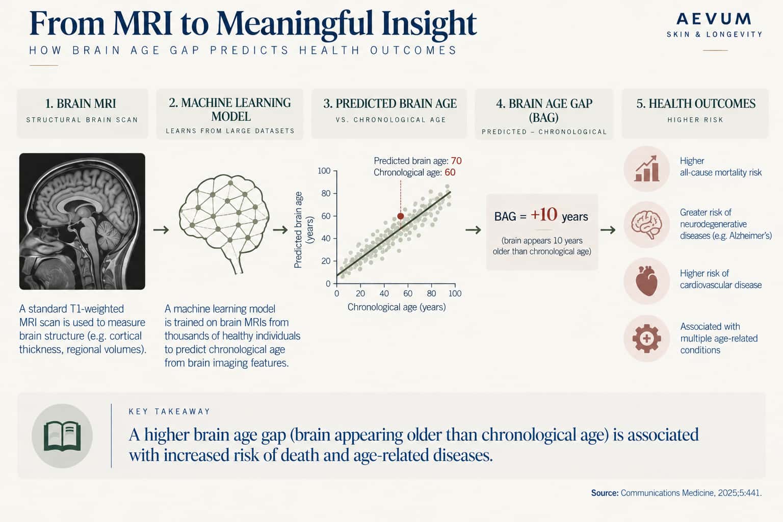
What "brain age" actually measures. Brain age is generated by training a machine-learning model on thousands of MRI scans from people of known chronological age, teaching it to recognize the structural patterns (volume, cortical thickness, white matter integrity) typical of each age. Once trained, the model can be shown a new scan and output a predicted age based purely on how the brain looks structurally. The "brain age gap" (BAG) is simply the difference between that predicted age and the person's actual chronological age — a positive gap means the brain looks structurally older than the person actually is; a negative gap means it looks younger.
The predictive power is large and validated across multiple independent cohorts. A large multi-cohort study — UK Biobank (n=38,967), the Alzheimer's Disease Neuroimaging Initiative (n=1,402), and the Parkinson's Progression Markers Initiative (n=1,182) — found that each additional year of brain age gap was associated with a 12% increase in all-cause mortality risk (median follow-up 3.4 years in the UK Biobank cohort). Comparing the extremes: people in the highest brain-age-gap quartile (brains looking oldest relative to actual age) had 2.36 times the mortality risk of those in the lowest quartile. The same gap was associated with a 16.5% higher Alzheimer's disease risk per year of gap, and measurable differences in processing speed and reaction time even in cognitively normal people, suggesting the metric picks up on subclinical decline before it becomes symptomatically obvious.
Brain age gap responds to modifiable factors. In the same study's highest-risk quartile, specific lifestyle factors were associated with measurably smaller brain age gaps: never smoking was linked to a 0.11-year reduction, moderate alcohol use to a 0.20-year reduction, and regular physical activity to a 0.14-year reduction. These are modest individual effects, but they reinforce a theme already established in this series (see our Neuroplasticity After 40 article): brain structure, even summarized as a single predictive number, isn't fixed — it responds to the same modifiable factors covered throughout this series.
What the research shows
Hippocampal volume works well for diagnosis, less well for early prediction. Separate from the whole-brain "brain age" metric, hippocampal volume specifically has been studied extensively as a diagnostic and predictive tool, since the hippocampus is one of the earliest and most consistently affected structures in Alzheimer's disease. A meta-analysis pooling 33 studies and 5,157 patients found hippocampal volumetry distinguishes Alzheimer's patients from healthy controls with solid accuracy (82% sensitivity, 87% specificity) — a genuinely useful diagnostic tool once the disease is established. But its accuracy drops meaningfully at the earlier, more clinically useful stage of distinguishing mild cognitive impairment from healthy aging (60% sensitivity, 75% specificity) — a real limitation worth being honest about, since the earlier stage is where the metric would be most valuable for early intervention. Interestingly, the entorhinal cortex — a structure closely connected to the hippocampus and among the very first regions affected in Alzheimer's pathology — performed somewhat better than the hippocampus alone in the same meta-analysis (88% sensitivity, 92% specificity for AD), suggesting the field's imaging targets are still evolving beyond hippocampal volume as the sole marker.
The honest limitation of any single volumetric marker. No single volume measurement, whether whole-brain "age" or hippocampal size specifically, functions as a stand-alone diagnostic test on its own — both are used clinically alongside cognitive testing, other imaging findings, and clinical history, not as isolated numbers. This connects directly to the reserve concept from our Cognitive Reserve article: two people with similar hippocampal volumes can present very differently clinically, which is part of why volumetric measures alone have real, quantified accuracy limits rather than functioning as perfect predictors.
Recommendations
- 1Don't over-interpret a single "brain age" number from a consumer or clinical scan in isolation
It's a genuinely useful population-level predictor with real statistical power, but an individual result carries meaningful uncertainty and should be interpreted alongside other clinical information, not as a standalone verdict.
- 2The lifestyle factors that measurably move brain age gap overlap heavily with the rest of this series
Physical activity, alcohol moderation, and not smoking are the specific factors with quantified associations here — a useful, concrete starting point rather than a vague "live healthier" recommendation.
- 3Hippocampal volume is a stronger diagnostic tool after symptoms appear than a predictive one before they do
This is useful context for interpreting a scan ordered after cognitive symptoms have already started, versus expecting it to reliably flag risk in someone who's currently asymptomatic.
- 4A "smaller than average" hippocampus alone, in someone with no cognitive symptoms, isn't a diagnosis
Given the meta-analysis's real false-positive/false-negative rates at the MCI stage specifically, an isolated volumetric finding in an otherwise asymptomatic person warrants context and follow-up, not alarm.
Practical notes
The consistent finding across both metrics in this article: brain structure measured on a scan is genuinely predictive of real outcomes, including mortality — but neither metric is a perfect crystal ball, and both are more useful as one input into a broader clinical picture than as a stand-alone verdict. Our upcoming Vascular Health and the Brain article covers one of the most consistently modifiable drivers behind these structural differences.
- Brain age gap as a predictive biomarker that links aging, lifestyle, and neuropsychiatric health. Communications Medicine, 2025;5:441.
- Diagnostic performance of hippocampal volumetry in Alzheimer's disease or mild cognitive impairment: a meta-analysis. European Radiology, 2022.
Have you ever had
a brain MRI or cognitive screening?
Take the free Aevum Protocol assessment to see how your cognitive longevity and 6 other longevity domains are performing — and get a personalised 90-day plan.
Take the Free Assessment →